Wojciech Krauze, PhD, awarded the ERC Starting Grant 2025
for the project Re.HOT — Reflection Holographic Tomography
Re.HOT — Reflection Holographic Tomography — is the project that has been awarded this year’s prestigious research grant of the European Research Council (ERC). This highly valued distinction was received by Wojciech Krauze, PhD, from the Faculty of Mechatronics at the Warsaw University of Technology. He is the second researcher from the Warsaw University of Technology to obtain an ERC grant in the Starting Grant category, and overall the third researcher from our University ever to become a laureate of a grant awarded by the European Research Council.
Reflection Holographic Tomography (Re.HOT) is a holographic tomography technique operating in reflection mode, comparable to a highly specialised microscope, in which a biological or medical sample is illuminated with coherent laser radiation from many different directions. Subsequently, using advanced computer algorithms, this set of projections is reconstructed and transformed into a three-dimensional map of the refractive index within the studied sample. Such a map carries detailed information about the internal microstructure of the investigated object.
This type of tomography is most frequently applied in the field of biology, where it is used to study living cell cultures, organoids, or thin tissue sections. A particularly important advantage is that there is no requirement for additional fluorescent staining. The major limitation of holographic tomography, however, is the fact that in order to obtain a high-quality, high-resolution image, the light source and the camera must be positioned on opposite sides of the sample under investigation.
— The purpose of the project is to enable holographic tomography to work effectively in reflection mode. Achieving this has been the aim of many research groups worldwide for many years. The reason is clear: it would allow researchers to obtain the same detailed information as in classical holographic tomography, but without the need to isolate and prepare patient samples. In our project we focus on the human cornea. On the one hand, the cornea is easier to work with because it is transparent. On the other hand, precise diagnostics of the cornea would open up a wide range of completely new applications in modern medicine — explains Wojciech Krauze, PhD, from the Faculty of Mechatronics at the Warsaw University of Technology, recipient of the ERC Starting Grant 2025 for the project entitled “Quantitative reflection holographic tomography for in-vivo analysis of biological specimens.”

The most fundamental research difficulty is that light which is reflected from tissue does not carry the same type of information as light transmitted directly through the sample. Because the range and quality of information is different, it is not possible to achieve identical results in reflection mode in a straightforward way. The greatest challenge, and simultaneously the central idea behind the Re.HOT project, is therefore to process the returning signal from the examined biological structure in such an intelligent and sophisticated manner that the obtained results are equivalent to those that would be produced if the tomography were performed in transmission mode.
A new quality in ophthalmology
The solution being developed by researchers at the Warsaw University of Technology will find applications primarily in the medical sector, and most directly in ophthalmology. At present, tools used for the assessment of corneal quality exist, but their practical application remains rather limited.
— If we succeed in reaching our intended objective, the project will result in a completely new measurement method that will complement and extend the set of currently available ophthalmic diagnostic tools. Until now, no researcher has been able to measure in a living patient the actual values of the refractive index in the cornea. A three-dimensional map of this coefficient will highlight locations with abnormal values, which in turn may allow for the very early detection of pathological changes in the cornea — emphasises Krauze, PhD.
Another potential advantage is associated with the development of a new generation of laser-based vision correction surgeries. Currently, the leading surgical techniques are still based on ablation — that is, the evaporation and removal of microscopic fragments of the corneal tissue. However, advanced research is being carried out on methods based on local modification of the refractive index of the cornea. These methods appear to be much safer for patients. What is missing, however, are control techniques that would allow ophthalmologists to determine and compare detailed refractive index maps both before and after the surgical procedure. The method developed by the research team at the Warsaw University of Technology may provide precisely such a solution.
Collaboration of experienced researchers
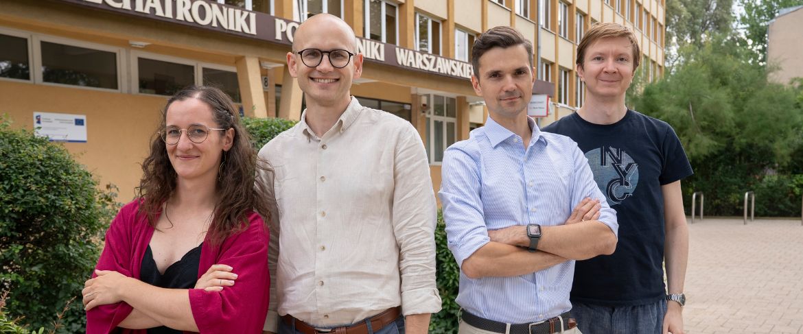
The Re.HOT project will be implemented by a team of eight researchers, led by Wojciech Krauze, PhD. Responsibility for the construction of the optical system has been entrusted to Arkadiusz Kuś, PhD. Doctoral candidate Martyna Mazur, MSc, will support Krauze, PhD, in the development of advanced data-processing algorithms. Maria Baczewska, PhD, will design a comprehensive measurement protocol and perform detailed data analysis, while Michał Ziemczonok, PhD, will use the technique of two-photon polymerisation to fabricate specialised phantoms on which the team will be able to test the initial version of the solution. All these researchers are staff members of the Faculty of Mechatronics at the Warsaw University of Technology, where the project will be implemented. In addition, the team plans to employ one student, one additional doctoral candidate, as well as an external medical expert — a researcher from outside the Warsaw University of Technology — who will support the researchers at the final stage of the project, when the system will be tested on a mouse cornea. The scientists talk about the project in the film.
For the purpose of preparing the project proposal, the team of WUT researchers conducted only simple proof-of-concept experiments. This does not mean, however, that they are starting “from scratch.” — Our team is composed of experienced scientists who have already participated in similar research projects. In addition, a useful point of departure in this work is the qtOCT method developed by us. This method represents a modification of classical OCT imaging. In our version, OCT operates in transmission mode and is therefore intended for examining biological samples isolated from the patient, for example placed on a microscope slide — adds Krauze, PhD.
As the researcher explains: — If we imagine light as consisting of photons travelling through tissue, some photons will travel quickly along short, unscattered paths, while others will scatter, wandering along much longer, circuitous routes. As a result, these scattered photons will exit the sample at a slightly later time. Scattered photons are always problematic, as they make it difficult to obtain high-quality images of the investigated sample. Our team has developed a technique that allows us to separate early photons from the later ones. Its fundamental limitation, however, is that it functions only in transmission mode, which means that the camera must be located on the side of the sample opposite the light source. Within the ERC project, our goal is to adapt this technique to reflection mode, which is a technically demanding and ambitious task.
The project can be divided into three principal stages:
- Construction of the optical system — including the design of the optical and mechanical layout, preparation of detailed specifications for all required components, their purchase and procurement, and finally the assembly and testing of the system.
- Development of advanced signal-processing algorithms — this stage will be carried out in parallel with the construction of the optical system.
- Validation of the complete system — first on micro-phantoms fabricated using two-photon polymerisation, and subsequently on biological samples, specifically the mouse cornea.
— Building the optical system itself will require a very large amount of work. It will be a relatively large and complex setup with numerous active components. Even the development of the electronics necessary to control and synchronise the functioning of all elements will be extremely challenging. We will need to plan the sequence of tasks very carefully, especially taking into account that under current legal and administrative regulations the purchase of scientific equipment may take many months. Another significant challenge will be working with the mouse cornea, which we have not previously dealt with — explains the laureate of the ERC grant.
Obtaining the ERC Starting Grant for the Re.HOT project gives the researchers the opportunity to carry out costly, long-term research: funding the salaries of the scientific team, purchasing all the required equipment (ranging from large optical tables to highly specialised system components), and above all concentrating fully on achieving ambitious scientific goals for the next five years. For the Warsaw University of Technology, this is not only a matter of prestige and international recognition, but also a way of systematically building its research capacity and strengthening its scientific position at the European level.
— For me personally, and for my entire research team, this award is both a source of great joy and also of considerable stress, because of the very high expectations involved — above all, our own expectations. For me it is confirmation that the research direction we have chosen is the right one. I would like to extend my deepest thanks to my wife for her constant support and invaluable logistical help in preparing the grant application, as well as to Professor Małgorzata Kujawińska, my mentor at the Warsaw University of Technology, who taught me how to conduct science at a truly world-class level. Without her guidance, I would never have had the courage or even the ambition to apply for European projects — concludes Wojciech Krauze, PhD.
The project “Quantitative reflection holographic tomography for in-vivo analysis of biological specimens” will be implemented at the Institute of Micromechanics and Photonics, Faculty of Mechatronics, Warsaw University of Technology. The funding allocated under the ERC Starting Grants amounts to EUR 1,499,775.